
This authoritative review article was published in Nature Reviews Drug Discovery in 2026, entitled Autophagy modulation in cancer. It was written by Emma Guilbaud, Kevin M. Ryan, Douglas R. Green and Lorenzo Galluzzi.
The central premise of this article is that autophagy exerts diametrically opposite effects at distinct stages of cancer development and progression:
In normal tissues: autophagy generally maintains cellular homeostasis and suppresses malignant transformation. In established tumours: autophagy frequently supports tumour progression and therapeutic resistance by enabling cancer cells to adapt to nutrient deprivation, hypoxia and therapeutic stress. Nevertheless, these effects are not absolute and exhibit strong context‑dependence.
At the immunological level: the impact of autophagy on tumour immune surveillance is highly context‑dependent; it can either suppress or promote anti‑tumour immunity.
Therefore, the article emphasises that there is no “one‑size‑fits‑all” strategy for autophagy modulation. Interventions must be tailored according to disease stage, tumour type and microenvironmental features.
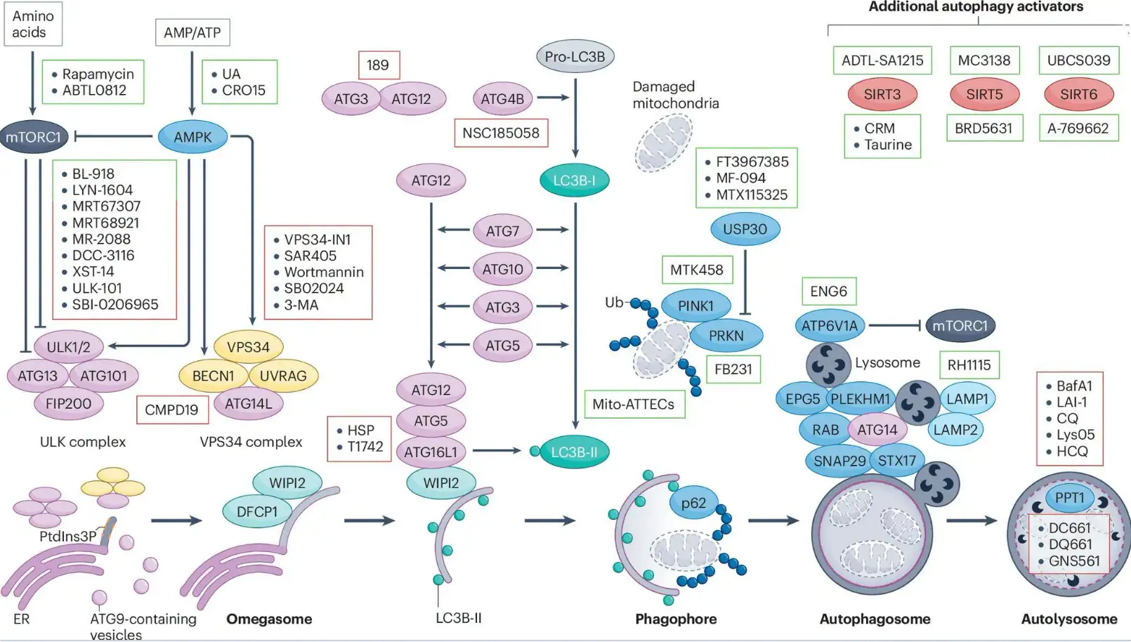
This review systematically summarises the core regulatory cascades of autophagy:
1. Initiation:
(1) mTORC1 is the major negative regulator of autophagy. It senses lysosomal amino‑acid levels and inhibits ULK1/2 and ATG13 via phosphorylation.
(2) AMPK is activated under energy deficiency (elevated AMP/ATP ratio). It promotes autophagy initiation by repressing mTORC1 and phosphorylating autophagy‑initiating factors such as ULK1.
(3) The VPS34 complex (comprising BECN1, UVRAG, ATG14L) generates PtdIns3P, driving phagophore expansion originating from ATG9‑containing vesicles.
2. Elongation & Closure:
Two ubiquitin‑like conjugation systems are essential:
(1) The ATG12‑ATG5‑ATG16L1 complex.
(2) Lipidation of LC3 (MAP1LC3): LC3‑I is processed by ATG4B, then lipidated via ATG7‑ and ATG3‑mediated ubiquitin‑like reactions to generate LC3‑II, which associates with autophagic membranes. Lipidated LC3‑II recognises and recruits ubiquitinated cargo by binding autophagy receptors such as p62/SQSTM1 for selective autophagy.
3. Maturation & Degradation:
(1) Autophagosomes fuse with lysosomes to form autolysosomes. This process requires SNARE proteins (STX17, SNAP29), RAB GTPases (RAB7A) and tethering factors (PLEKHM1, EPG5).
(2) Lysosomal acidification and hydrolase activity (e.g. PPT1) are indispensable for substrate degradation.
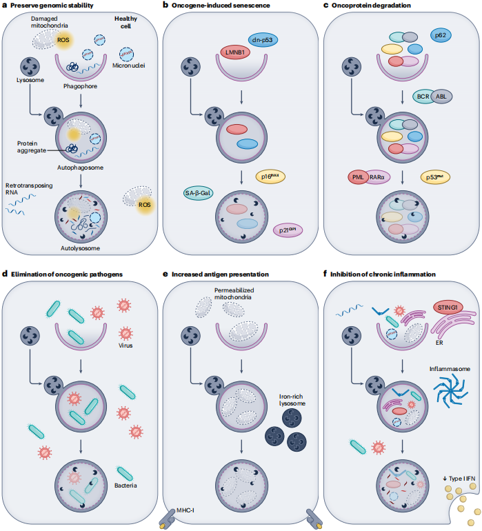
In normal cells, autophagy restrains malignant transformation via six major mechanisms:
| Mechanism | Description |
|---|---|
| a. Maintain genomic stability | Eliminate ROS‑producing damaged mitochondria, protein aggregates, micronuclei and retrotransposon RNAs |
| b. Oncogene‑induced senescence | Autophagy participates in oncogene‑driven cellular senescence, linked to LMNB1 degradation and senescence hallmarks including p16/p21 and SA‑β‑Gal |
| c. Degradation of oncogenic proteins | Selective degradation of mutant p53, p62, BCR‑ABL, PML‑RARα and others |
| d. Clearance of oncogenic pathogens | Remove oncogenic bacteria and viruses through xenophagy |
| e. Boost antigen presentation | Mitophagy promotes lysosomal iron accumulation and up‑regulates MHC‑I expression, favouring immune recognition of nascent malignant cells |
| f. Suppress chronic inflammation | Degrade endogenous activators of pattern‑recognition receptors (micronuclei, mitochondrial DNA, STING1 etc.) and dampen tumour‑promoting inflammation |
Heterozygous or tissue‑specific deletion of multiple autophagy‑related genes including Becn1, Ambra1, Atg5/Atg7 can drive endogenous tumour formation in certain mouse models. However, such effects exhibit strong tissue‑ and genetic‑context dependency; autophagy‑related genes cannot be simply classified as canonical tumour suppressors.
Notably, under specific genetic backgrounds, Becn1 heterozygous loss may exert phenotypes opposite to its canonical tumour‑suppressive function. For instance, it blocks tumour progression in the Tsc2+/- model, implying that BECN1 possesses autophagy‑independent biological functions.
Non‑pharmacological interventions: caloric restriction, physical exercise, fasting‑mimicking diet (FMD). These interventions exert anti‑tumour or sensitising effects in multiple animal models, yet definitive mechanistic evidence proving that benefits are primarily autophagy‑dependent remains insufficient.
Potential pharmacological interventions: Rapamycin and partial caloric‑restriction mimetics (e.g. α‑ketoglutarate, resveratrol, aspirin) may confer anti‑tumour activities by modulating autophagy and metabolic pathways; however their effects are not necessarily fully autophagy‑dependent.
Key challenges: Measuring autophagic flux in human tissues is technically difficult. Most available studies rely on static markers rather than dynamic flux readouts. Assessing only static markers such as LC3B‑II, LC3 puncta or p62/SQSTM1 cannot reliably reflect actual autophagic flux. Associations between autophagic activity and therapeutic benefits therefore require combined dynamic‑flux and functional evidence.
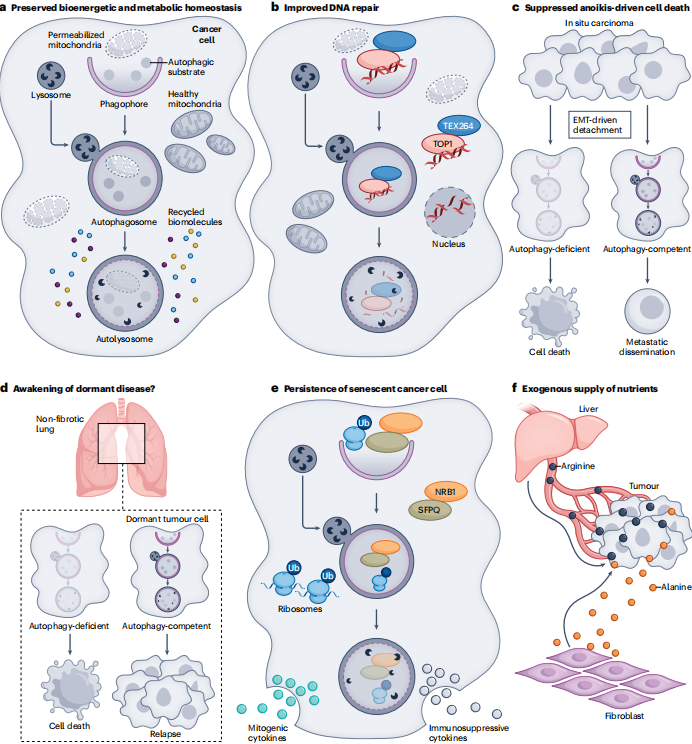
In established tumours, autophagy sustains tumour progression and therapeutic resistance via the following mechanisms:
| Mechanism | Description |
|---|---|
| a. Sustain bioenergetic homeostasis | Recycle macromolecules under nutrient‑oxygen deprivation to maintain metabolic homeostasis |
| b. Facilitate DNA repair | Remove DNA‑damage foci via selective autophagy mediated by TEX264 and other factors |
| c. Resist anoikis | Support survival of detached cancer cells during EMT and promote metastasis |
| d. Maintain dormant tumour cells | Support survival of cancer stem cells and disseminated dormant tumour cells; influence reactivation and relapse, yet subsequent expansion is highly context‑dependent |
| e. Persistence of senescent cancer cells | Promote survival of senescent cancer cells that continuously secrete pro‑tumorigenic and immunosuppressive factors |
| f. Exogenous nutrient supply | Tumour microenvironment (hepatocytes, fibroblasts) provides alanine, arginine and other metabolites through autophagy |
Deletion of Atg5/Atg7 markedly impairs tumour progression in KRAS/BRAF‑driven lung and pancreatic cancer mouse models.
Nevertheless, context‑dependent paradox exists: under pulmonary‑fibrosis background, autophagy inhibition may aggravate metastatic burden, partially caused by accumulation of NBR1 and PFKFB3 plus re‑awakening of dormant tumour cells.
Of note, loss of Ambra1 can produce divergent or even opposite phenotypes at distinct tumour stages, further highlighting context dependency of autophagy regulation.
Clinical translation of CQ/HCQ has yielded overall unsatisfactory results. Multiple phase‑II studies show null or marginal clinical benefit. This cannot be simply interpreted as “autophagy inhibition being ineffective”, because these agents possess substantial off‑target effects.
Reasons for limited clinical efficacy:
- These agents are highly non‑specific with modest anti‑cancer potency (EC50 ranging 10‑50 μM).
- Autophagy inhibition can trigger compensatory KEAP1‑NRF2 activation, enhancing macropinocytosis and proteasomal degradation.
- Many anti‑tumour effects may arise from autophagy‑independent mechanisms such as vascular normalisation.
This represents the most complex section of this review, given that autophagy‑immune crosstalk is profoundly context‑dependent.
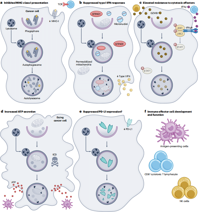
Autophagy in cancer cells can dampen anti‑tumour immunity via these mechanisms:
a. Suppress MHC‑I antigen presentation: autophagy degrades MHC‑I molecules and reduces cancer‑cell visibility to CD8+ cytotoxic T‑lymphocytes.
b. Repress type‑I interferon responses: degrade immunostimulatory structures including STING1, micronuclei and permeabilised mitochondria, thus blunting type‑I‑IFN / cGAS‑STING signalling.
c. Increase resistance to cytotoxic effectors: degrade cytosolic GZMB and reduce tumour‑cell responsiveness to IFNγ, TNF and other inflammatory‑cytokine‑mediated cytotoxicity.
Genetic evidence:
- Atg16l1 deletion sensitises colorectal cancer cells to IFNγ and TNF.
- Knock‑down of BECN1/ATG5/p62 sensitises melanoma to cytotoxic T‑lymphocyte killing.
- Dominant‑negative ATG4B restores MHC‑I presentation and potentiates PD‑1‑blockade efficacy.
Pharmacological evidence:
- Lysosomal inhibitors CQ/HCQ as well as more selective autophagy‑pathway inhibitors (e.g. SB02024) can enhance ICI responses in selected tumour models.
- The PPT1‑targeting inhibitor DC661 improves PD‑1‑blockade responses in some pre‑clinical tumour models, partially linked to restored type‑I‑IFN / STING signalling, though the precise molecular mechanism remains incompletely characterised.
Paradoxically, autophagy can promote anti‑tumour immunity under certain circumstances:
d. ATP release during immunogenic cell death (ICD): autophagy is critical for ATP release upon chemotherapy‑ or radiotherapy‑triggered ICD. In syngeneic colorectal‑cancer mouse models, tumour‑cell‑specific deletion of Atg5/Atg7 completely abolishes mitoxantrone therapeutic efficacy, demonstrating tumour‑cell autophagy is essential for certain ICD‑based chemotherapies.
e. PD‑L1 degradation: accumulating evidence suggests autophagy may target PD‑L1 for lysosomal degradation (requires further validation).
f. Immune‑effector‑cell development: autophagy is essential for proper development and function of NK cells, CD8+ T‑cells and dendritic cells.
In myeloid cells, LAP (non‑canonical autophagy) fosters tumour immune tolerance by suppressing type‑I‑IFN responses.
Deletion of Atg5/Atg7/Atg16l1 (perturbs canonical autophagy plus LAP) or Rubcn/Nox2 (selectively perturbs LAP) restricts tumour progression in pre‑clinical models.
This indicates systemic autophagy modulators influence not only canonical autophagy but also LAP, thereby reshaping the tumour immune microenvironment.
This landmark review synthesises current knowledge linking autophagy and cancer. Its major contribution is a systematic elaboration of the context‑dependent dual roles of autophagy, breaking oversimplified viewpoints that “autophagy is universally good or bad”.
It emphasises the significance of immune microenvironment: systemic autophagy modulation affects both tumour cells and host immune cells simultaneously.
It constructs a complete research‑to‑clinical‑translation chain: highlighting promising next‑generation selective agents (e.g. ULK1 inhibitors, PPT1 inhibitors), while honestly acknowledging practical obstacles including lack of reliable biomarkers and compensatory signalling circuits.
Autophagy inside tumour cells, immune cells and other host compartments can exert divergent, even opposing biological outcomes. Simple systemic autophagy activation or suppression strategies cannot be universally applied.
Reference
Guilbaud E, Ryan KM, Green DR, Galluzzi L. Autophagy modulation in cancer. Nat Rev Drug Discov. 2026;25:701‑722.
| Target | Catalog# | Product Name | Reactivity | Application |
|---|---|---|---|---|
| ULK1 | AMRe83955 | ULK1 Rabbit Monoclonal Antibody | Human,Mouse,Rat | WB,IHC,ICC/IF,ICC |
| ULK2 | AMM81619 | ULK2 Mouse Monoclonal Antibody | Human,Mouse,Rat | WB,ELISA,FC |
| ATG13 | AMRe85134 | ATG13 Rabbit Monoclonal Antibody | Human,Mouse,Rat | WB,IP |
| RB1CC1 | AMM82303 | RB1CC1 Mouse Monoclonal Antibody | Human | ELISA |
| AMPK α1 | AMRe21607 | AMPK α1 Rabbit Monoclonal Antibody | Human,Mouse,Rat | WB,IHC,ICC/IF,ELISA,IP |
| AMPK beta 1 | AMRe06843 | AMPK beta 1 (3Y15) Rabbit Monoclonal Antibody | Human,Mouse,Rat | WB,IHC,ICC/IF,FC,IP |
| AMPK gamma 1 | AMRe86981 | AMPK gamma 1 Rabbit Monoclonal Antibody | Human, Mouse, Rat | WB,FC,IP |
| VPS34 | AMRe87144 | VPS34 Rabbit Monoclonal Antibody | Human,Mouse,Rat | WB,IHC |
| BECN1 | AMM80962 | BECN1 Mouse Monoclonal Antibody | Human,Mouse | WB,IHC,ELISA,FC |
| ATG14L | AMM81601 | ATG14L Mouse Monoclonal Antibody | Human | IHC,ELISA |
| WIPI2 | APRab55662 | WIPI2 Rabbit Polyclonal Antibody | Human,Mouse,Rat | IHC,IF |
| DFCP1/ZFYV1 | APRab79429 | ZFYV1 Rabbit Polyclonal Antibody | Human,Mouse | WB |
| ATG3 | AMRe21180 | ATG3 Rabbit Monoclonal Antibody | Human,Mouse,Rat | WB,IHC,IF,ELISA |
| Apg12 | AMRe83957 | Apg12(Atg12) Rabbit Monoclonal Antibody | Human | WB,IHC,ICC/IF,ICC,FC,IP |
| LC3B | AMRe21536 | LC3B Rabbit Monoclonal Antibody | Human,Mouse,Rat | WB,IHC,ICC/IF,ELISA,IP |
| ATG4B | AMM82334 | ATG4B Mouse Monoclonal Antibody | Human, Mouse | WB,ELISA,FC |
| ATG5 | AMRe21348 | ATG5 Rabbit Monoclonal Antibody | Human,Mouse,Rat,Chicken | WB,IHC,IF,IP,ELISA |
| ATG16L1 | AMRe21136 | ATG16L1 Rabbit Monoclonal Antibody | Human,Mouse,Rat | WB,IHC,ICC/IF,ELISA,IP |
| ATG7 | AMRe21471 | ATG7 Rabbit Monoclonal Antibody | Human,Mouse,Rat | WB,IHC,ICC/IF,ELISA,IP |
| ATG10 | APRab07282 | ATG10 Rabbit Polyclonal Antibody | Human,Rat,Mouse | IHC,ICC/IF,ELISA |
| USP30 | APRab19673 | USP30 Rabbit Polyclonal Antibody | Human,Mouse,Rat | WB,ELISA |
| PINK1 | AMM85907 | PINK1 Mouse Monoclonal Antibody | Human, Mouse | WB,IHC |
| PRKN | AMM82302 | PRKN Mouse Monoclonal Antibody | Human | ELISA,FC |
| p62/SQSTM1 | AMRe15658 | p62/SQSTM1 (12N5) Rabbit Monoclonal Antibody | Human,Mouse,Rat | WB,IHC,ICC/IF,FC,IP |
| ATP6V1A | AMRe85328 | ATP6V1A Rabbit Monoclonal Antibody | Human,Mouse,Rat | WB,IHC,IP |
| EPG5 | APRab10509 | EPG5 Rabbit Polyclonal Antibody | Human,Rat,Mouse | WB,IHC,ICC/IF |
| PLEKHM1 | APRab57967 | PLEKHM1 Rabbit Polyclonal Antibody | Human,Mouse,Rat | WB |
| SNAP29 | AMRe18048 | SNAP29 (10N2) Rabbit Monoclonal Antibody | Human,Mouse,Rat | WB,ICC/IF,FC,IP |
| STX17 | AMM83641 | STX17 Mouse Monoclonal Antibody | Human | WB,IHC |
| LAMP1 | AMRe87845 | LAMP1 Rabbit Monoclonal Antibody | Mouse | WB,IHC,ICC/IF,FC,IP |
| LAMP2 | AMRe01588 | LAMP2 Rabbit Monoclonal Antibody | Human,Mouse,Rat | WB,IHC,IF,ELISA |
| PPT1 | AMM85956 | PPT1 Mouse Monoclonal Antibody | Human | WB,IHC,FC |
| SIRT3 | AMRe21475 | SIRT3 Rabbit Monoclonal Antibody | Human,Mouse,Rat | WB,IHC,ICC/IF,ELISA,IP |
| SIRT5 | AMRe83856 | SIRT5 Rabbit Monoclonal Antibody | Human,Mouse | WB |
| SIRT6 | AMRe17919 | SIRT6 (10O9) Rabbit Monoclonal Antibody | Human,Mouse,Rat | WB,IHC,ICC/IF,IP |
Related Products